


Tomography 2024, 10(5), 761-772; https://doi.org/10.3390/tomography10050058 - 15 May 2024
Abstract
Lymphadenectomy represents a fundamental step in the staging and treatment of non-small cell lung cancer (NSCLC). To date, the extension of lymphadenectomy in early-stage NSCLC is a debated topic due to its possible complications. The detection of sentinel lymph nodes (SLNs) is a
[...] Read more.
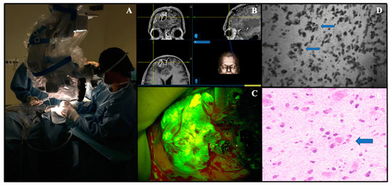
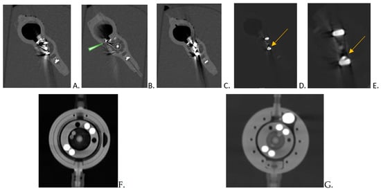
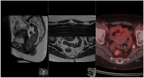
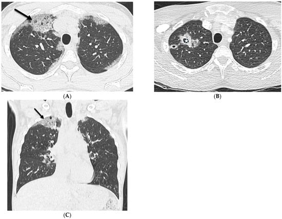
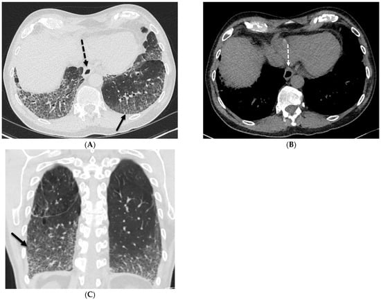
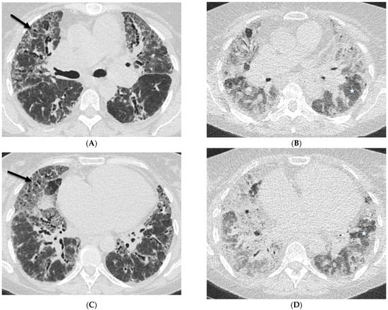
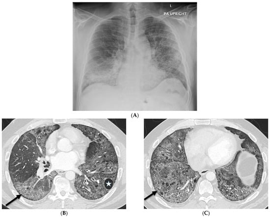
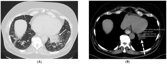
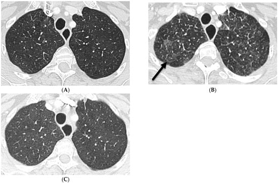
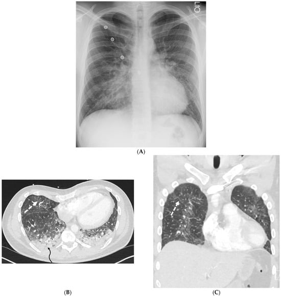
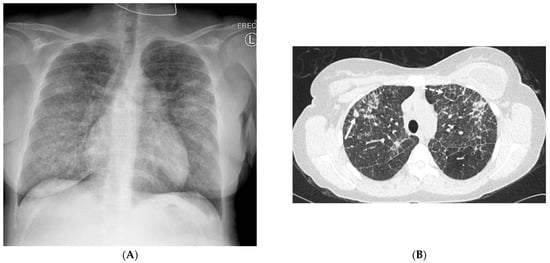
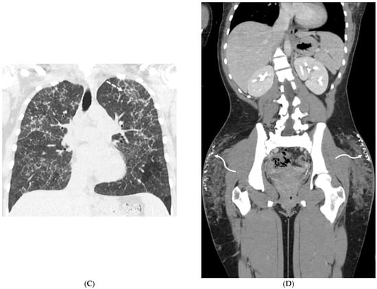
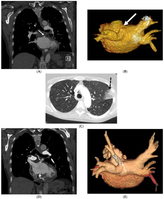
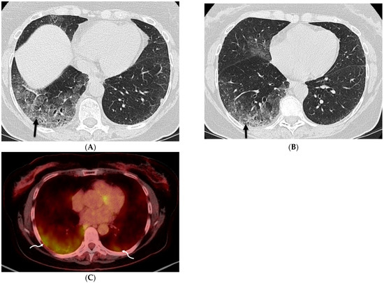
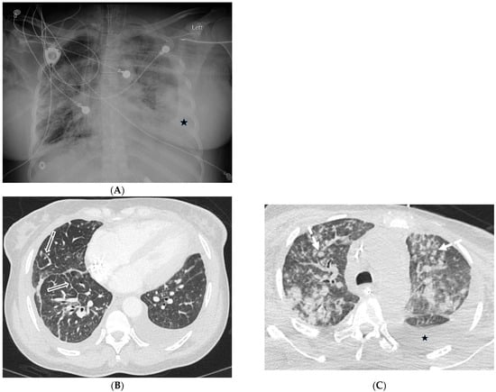
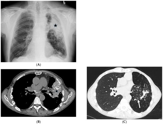
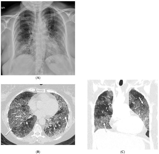
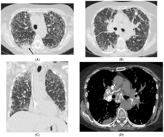
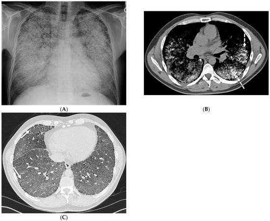
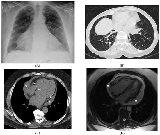
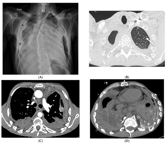
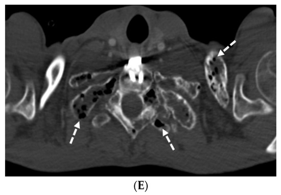
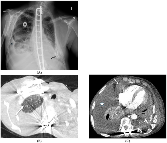
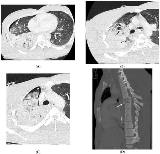
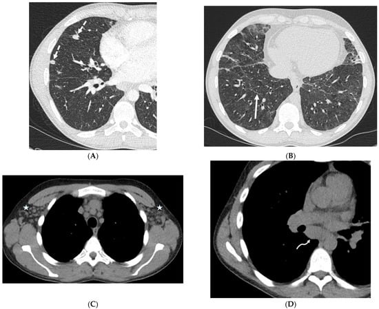
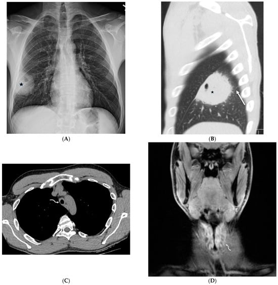
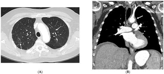
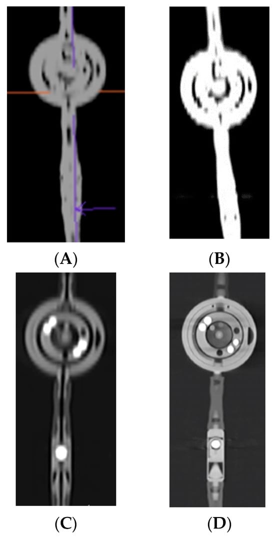
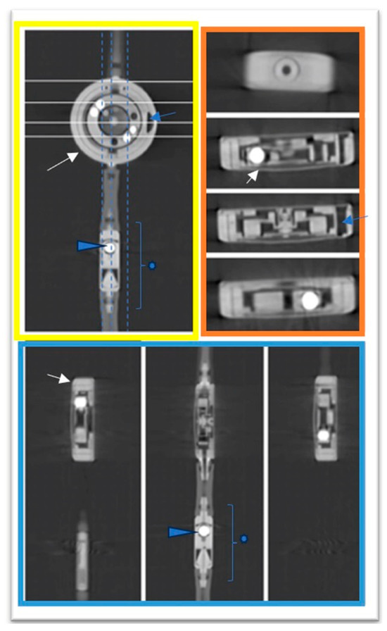
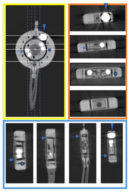
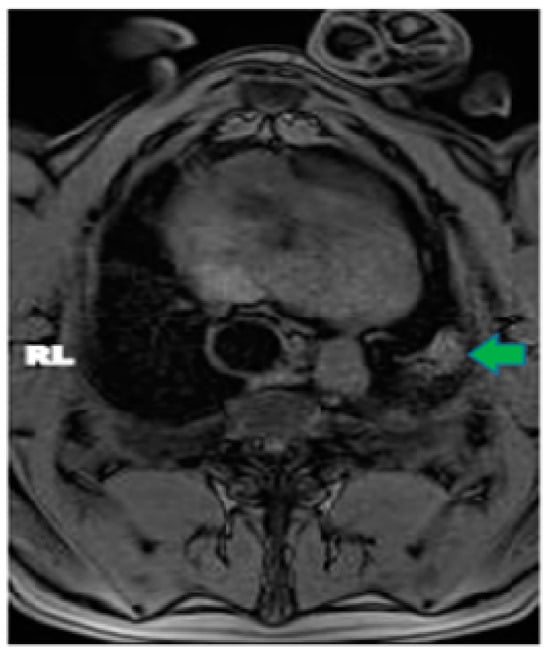
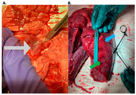
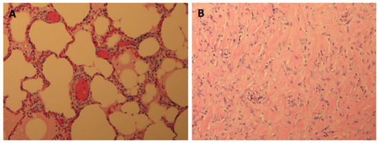
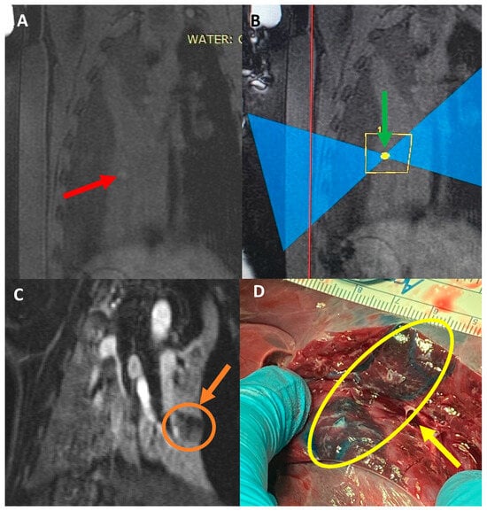
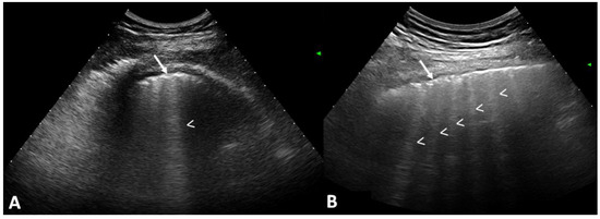
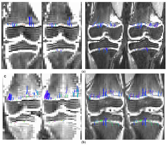
Lymphadenectomy represents a fundamental step in the staging and treatment of non-small cell lung cancer (NSCLC). To date, the extension of lymphadenectomy in early-stage NSCLC is a debated topic due to its possible complications. The detection of sentinel lymph nodes (SLNs) is a strategy that can improve the selection of patients in which a more extended lymphadenectomy is necessary. This pilot study aimed to refine lymph nodal staging in early-stage NSCLC patients who underwent robotic lung resection through the application of innovative intraoperative sentinel lymph node (SLN) identification and the pathological evaluation using one-step nucleic acid amplification (OSNA). Clinical N0 NSCLC patients planning to undergo robotic lung resection were selected. The day before surgery, all patients underwent radionuclide computed tomography (CT)-guided marking of the primary lung lesion and subsequently Single Photon Emission Computed Tomography (SPECT) to identify tracer migration and, consequently, the area with higher radioactivity. On the day of surgery, the lymph nodal radioactivity was detected intraoperatively using a gamma camera. SLN was defined as the lymph node with the highest numerical value of radioactivity. The OSNA amplification, detecting the mRNA of CK19, was used for the detection of nodal metastases in the lymph nodes, including SLN. From March to July 2021, a total of 8 patients (3 female; 5 male), with a mean age of 66 years (range 48–77), were enrolled in the study. No complications relating to the CT-guided marking or preoperative SPECT were found. An average of 5.3 lymph nodal stations were examined (range 2–8). N2 positivity was found in 3 out of 8 patients (37.5%). Consequently, pathological examination of lymph nodes with OSNA resulted in three upstages from the clinical IB stage to pathological IIIA stage. Moreover, in 1 patient (18%) with nodal upstaging, a positive node was intraoperatively identified as SLN. Comparing this protocol to the usual practice, no difference was found in terms of the operating time, conversion rate, and complication rate. Our preliminary experience suggests that sentinel lymph node detection, in association with the accurate pathological staging of cN0 patients achieved using OSNA, is safe and effective in the identification of metastasis, which is usually undetected by standard diagnostic methods.
Full article
(This article belongs to the Section Cancer Imaging)
►
Show Figures

Figure 1


